About Me

I am a Ph.D. candidate in Computer Science and Technology at Harbin Institute of Technology, Shenzhen, advised by Prof. Yongbing Zhang. I am also a government-sponsored visiting Ph.D. student at TU Dresden, advised by Prof. Jakob Nikolas Kather.

My research interests include multimodal medical AI, AI agents, and computational pathology. I study how learning systems can connect gigapixel pathology, clinical information, medical imaging, and molecular measurements while keeping their predictions interpretable and clinically reviewable.

My recent work spans virtual multiplex immunofluorescence and spatial immune analysis, smartphone-based intraoperative assessment, weakly supervised whole-slide learning, and agentic reasoning over gigapixel pathology images. I am always open to exchanging ideas and would be delighted to connect, so please feel free to email me.

Related work has appeared in Nature Genetics, The Lancet Digital Health, IEEE Transactions on Medical Imaging, Medical Image Analysis, ICLR, and ICML.

📢📢 Current focus: multimodal medical AI, AI agents, and computational pathology. I am seeking full-time opportunities through the 2027 graduate recruitment cycle. Contact: cailh@stu.hit.edu.cn · CV.

  • Multimodal Medical AIPathology · clinical data · imaging · genomics
  • AI AgentsEvidence retrieval · cross-scale reasoning · interpretable decisions
  • Computational PathologyWeak supervision · spatial analysis · biomarker discovery

News

  • STEP was accepted by Nature Genetics.
  • Released PathAgentBench and PathScale-R1, a benchmark and a training framework for cross-scale pathology reasoning.
  • SuRImage was accepted by The Lancet Digital Health.
  • Released HCCExplorer for virtual multiplex immunofluorescence and spatial immune analysis.
  • Released PathReasoner-R1 for knowledge-guided pathology reasoning.
  • AttriMIL was published in Medical Image Analysis.

Selected Publications

Full list: Google Scholar Google Scholar citations: 620 h-index 14 · i10-index 15

🧬 Spatial Pathology & Multimodal Cancer Analysis

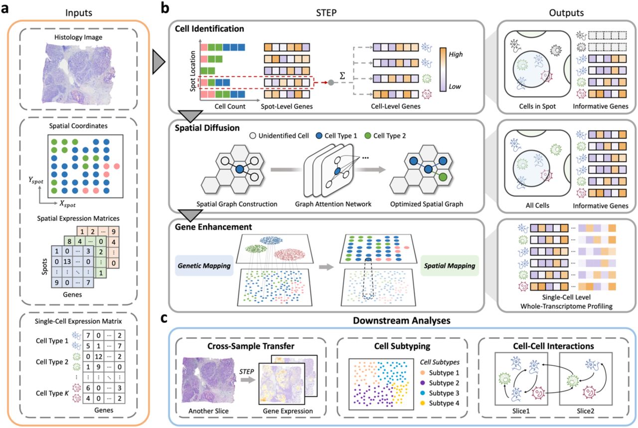
Nature Genetics 2026 STEP framework for single-cell whole-transcriptome spatial analysis
Co-first AuthorAccepted 2026

STEP: Deciphering Spatial Atlas at Single-Cell Level with Whole-Transcriptome Coverage

Zheqi Hu, Zirui Zhu, Linghan Cai, et al.

  • Question: Can spatial transcriptomics achieve both single-cell resolution and whole-transcriptome coverage while preserving tissue spatial organization?
  • Approach: STEP combines probabilistic modeling, deep learning, and histological nuclear features through Cell Identification, Spatial Diffusion, and Gene Enhancement.
  • Finding: The framework reconstructs cell types and transcriptome-wide expression, supports cross-sample transfer, and enables spatially resolved analysis of cell-cell interactions.
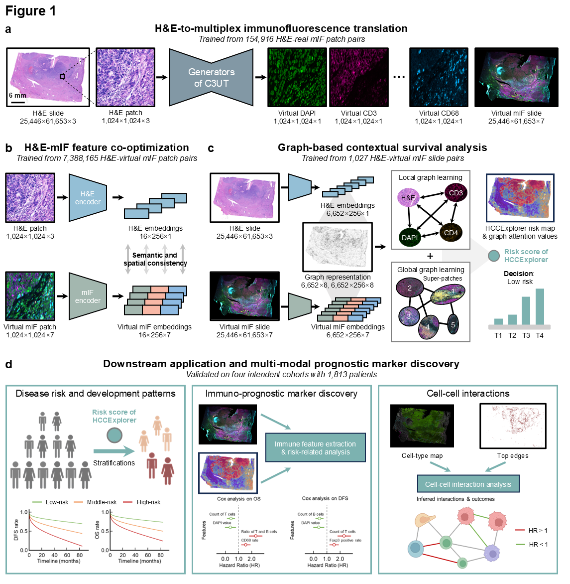
Under review · Science Advances HCCExplorer framework for virtual multiplex immunofluorescence and spatial survival analysis
First Author2026

Transforming Histology into Virtual Multiplex Immunofluorescence to Decode Prognostic Spatial Immunity in Hepatocellular Carcinoma

Linghan Cai, Songhan Jiang, Junhao Liang, et al.

  • Question: Can routine H&E slides recover multiplex immune phenotypes and prognostic spatial organization without physical multiplex staining?
  • Approach: HCCExplorer combines cell-consistent H&E-to-virtual-mIF translation, multimodal feature co-optimization, and graph-based contextual survival learning.
  • Finding: Across 1,813 patients from four centers, the framework achieved a reported C-index of 0.71 and identified a macrophage-associated containment niche at the invasion frontier.
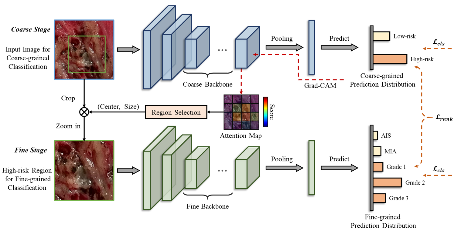
Lancet Digital Health 2026 SuRImage coarse-to-fine model framework for surgical resection image analysis
Co-first Author20261 citation

Deep Learning Model for Pathological Invasiveness Prediction Using Smartphone-based Surgical Resection Images in Clinical Stage IA Lung Adenocarcinoma (SuRImage): a Prospective, Multicentric, Diagnostic Study

Lintong Yao, Linghan Cai, Maotao Weng, et al.

  • Question: Can smartphone photographs of fresh surgical specimens support rapid intraoperative assessment of pathological invasiveness in stage IA lung adenocarcinoma?
  • Approach: SuRImage uses a coarse-to-fine visual model for invasiveness diagnosis, suspicious-region identification, and subtype classification in a prospective multicenter study.
  • Finding: The study demonstrates that macroscopic resection images contain learnable morphological cues that can complement time-sensitive intraoperative decision-making.

🤖 Pathology Foundation Models & Agents

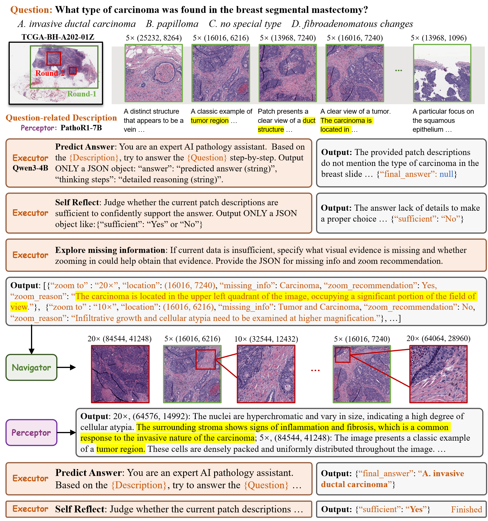
ECCV 2026 PathAgent iterative reasoning, navigation, and evidence retrieval workflow
Co-first AuthorECCV 202618 citations

PathAgent: Toward Interpretable Analysis of Whole-slide Pathology Images via Large Language Model-based Agentic Reasoning

Jingyun Chen, Linghan Cai, Zhikang Wang, et al.

  • Question: Can an AI agent actively inspect a gigapixel whole-slide image and make its diagnostic evidence traceable?
  • Approach: PathAgent coordinates an Executor, Navigator, and Perceptor to plan multi-step analysis, select magnification, retrieve evidence, and revise conclusions.
  • Finding: A training-free agentic workflow produced reviewable reasoning paths while retaining competitive performance with off-the-shelf pathology models.
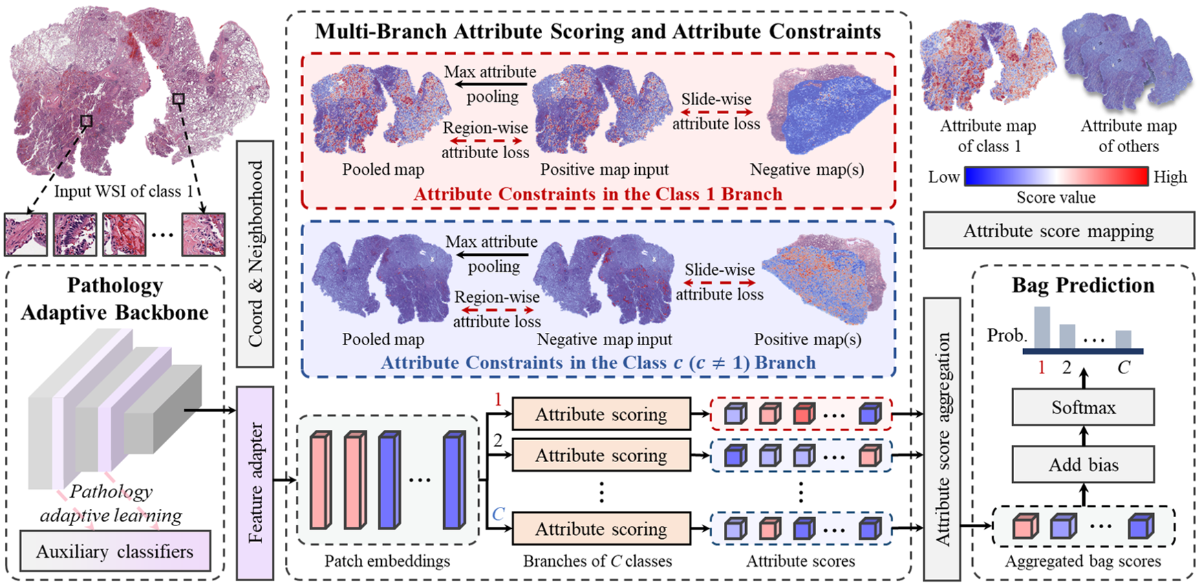
MedIA 2025 AttriMIL architecture with attribute scoring branches and constraints
First AuthorMedIA 202546 citations

AttriMIL: Revisiting Attention-based Multiple Instance Learning for Whole-slide Pathological Image Classification from a Perspective of Instance Attributes

Linghan Cai, Shenjin Huang, Ye Zhang, Jinpeng Lu, Yongbing Zhang

  • Question: Do attention scores faithfully measure the pathological attributes of individual tissue instances in weakly supervised slide classification?
  • Approach: AttriMIL introduces multi-branch attribute scoring, region- and slide-level constraints, and pathology-adaptive feature learning.
  • Finding: The method achieved state-of-the-art results across four benchmarks and improved localization of disease-positive regions, including slides with sparse targets.

⚡ Efficient & Data-efficient Medical Vision

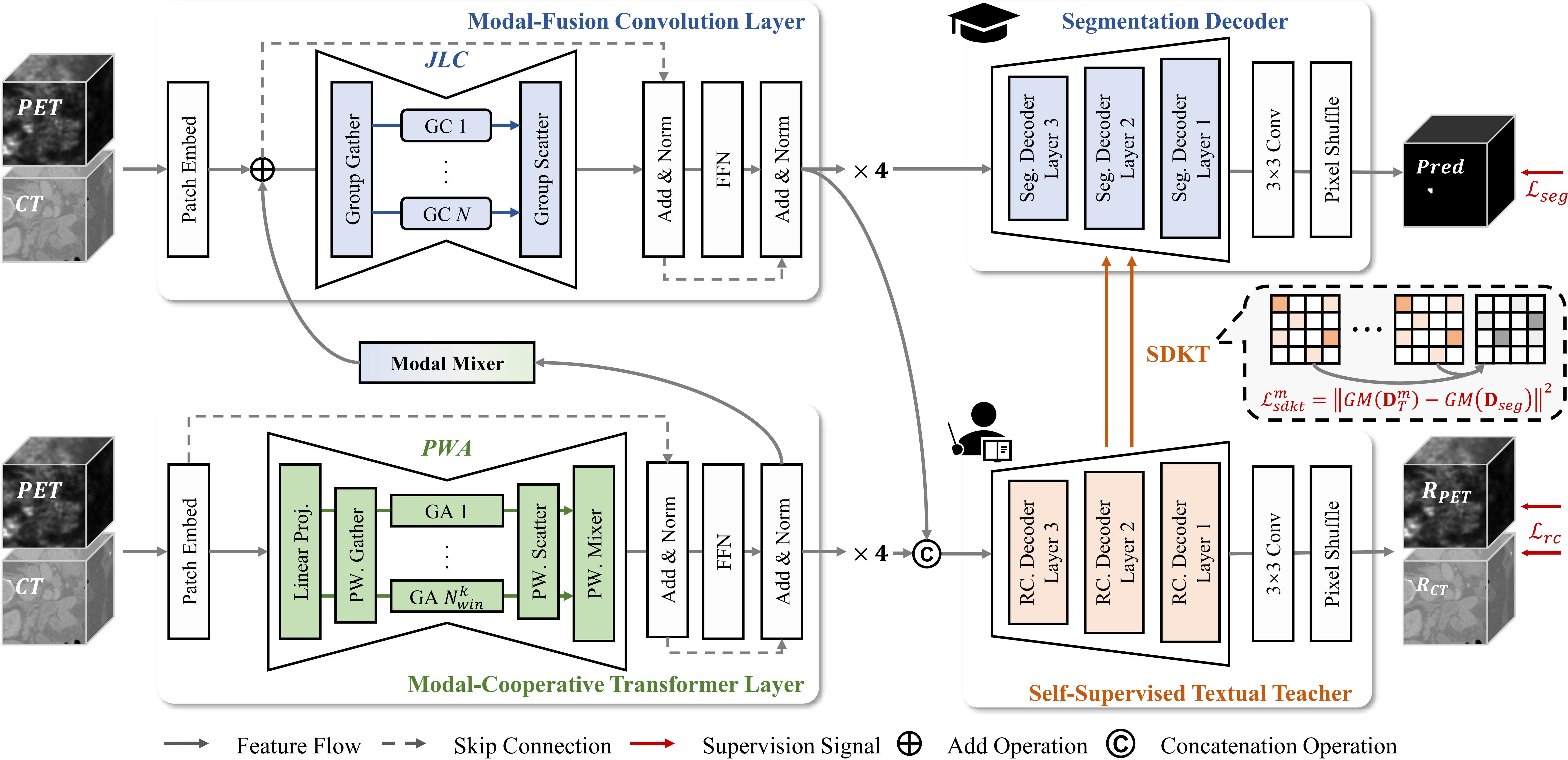
ICLR 2026 VeloxSeg architecture with modal-fusion convolution and modal-cooperative transformer layers
Co-first AuthorICLR 20261 citation

Johnson-Lindenstrauss Lemma Guided Network for Efficient 3D Medical Segmentation

Jinpeng Lu, Linghan Cai, Yinda Chen, et al.

  • Question: Can multimodal 3D segmentation remain accurate and robust without the computational cost of large volumetric networks?
  • Approach: VeloxSeg combines Johnson-Lindenstrauss-guided convolution, paired-window attention, modal mixing, and self-distilled dual decoders.
  • Finding: The reported model uses 1.66M parameters and 1.79G FLOPs while maintaining competitive segmentation across PET/CT and MRI benchmarks.
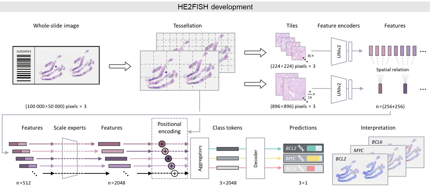
Under review · npj Digital Medicine HE2FISH multiscale expert framework for gene rearrangement prediction
First Author2026

Data-efficient Deep Learning for Gene Rearrangement Status Prediction in Diffuse Large B-cell Lymphoma

Linghan Cai, et al.

  • Question: Can routine H&E slides provide a data-efficient screening signal for BCL2, BCL6, and MYC rearrangements in diffuse large B-cell lymphoma?
  • Approach: HE2FISH extracts 20× and 5× UNI2-h features and integrates them with multiscale experts, positional encoding, and task-specific prediction tokens.
  • Finding: The framework supports all three rearrangement tasks and produces interpretable attention maps that localize morphology associated with each prediction.

Research

🧬 Multimodal Medical AI

Connecting complementary clinical, imaging, pathological, and molecular evidence.

I develop models that integrate clinical variables, radiology, histopathology, and gene expression for cancer diagnosis and prognosis. The central challenge is not simply feature fusion, but determining which modality contributes actionable evidence for a patient-level decision.

SuRImage studies smartphone-based intraoperative assessment, while HCCExplorer connects routine H&E, virtual multiplex immunofluorescence, graph learning, and spatial immune biomarkers.

🔬 Computational Pathology

Learning from gigapixel slides under weak supervision while preserving interpretable morphology.

My work covers whole-slide preprocessing, multiple instance learning, nuclei analysis, spatial modeling, and biomarker discovery. I am especially interested in the relationship between slide-level predictions and the local tissue attributes that support them.

AttriMIL revisits attention-based MIL through instance attributes, and related projects study weakly supervised nuclei segmentation, spatial transcriptomics, and pathology-genomics survival analysis.

🤖 AI Agents for Pathology

Turning static prediction into active, cross-scale, evidence-grounded analysis.

I build agentic systems that navigate whole-slide images, select magnification, retrieve morphological evidence, and expose a reviewable reasoning process. This shifts pathology AI from one-shot classification toward iterative analysis that can be inspected by human experts.

PathAgent, PathReasoner-R1, and PathScale-R1 explore interactive slide navigation, structured reasoning, and cross-scale pathology evaluation.

⚡ Earlier Foundation: Efficient Medical Vision

Efficient and data-efficient learning for segmentation and molecular prediction.

My earlier work develops lightweight and robust image-analysis models for polyp segmentation, multimodal 3D medical segmentation, and pathology-based molecular screening. These projects provide the efficiency foundations for deploying richer multimodal and agentic systems.

Research Experience

Multimodal Medical Data Fusion and Analysis

Project Lead. Integrating clinical information, radiomics, pathology, and gene expression for precise cancer diagnosis, prognosis, and spatial biomarker discovery.

AI-driven Computational Pathology

Project Lead. Building standardized digital-pathology pipelines and learning methods for weak supervision, nuclei analysis, whole-slide reasoning, and interpretable biomarker extraction.

Efficient and Robust Image Analysis

Core Contributor. Designing efficient deep-learning architectures for polyp segmentation, multimodal 3D segmentation, and data-efficient pathology analysis.

Education

Harbin Institute of Technology, Shenzhen

Ph.D. in Computer Science and Technology. Advisor: Prof. Yongbing Zhang.

GPA: 3.45 / 4.00 (top 20%). Research focus: computational pathology and multi-omics analysis. Recipient of a First-Class Scholarship.

TU Dresden

Government-sponsored Visiting Ph.D. Student in Computer Science and Technology. Advisor: Prof. Jakob Nikolas Kather.

Beihang University

M.S. in Circuits and Systems.

GPA: 3.80 / 4.00 (top 10%, 19 / 193). Research focus: biomedical signal processing. First Prize in the Beijing Graduate Electronic Design Contest and National Third Prize.

China Agricultural University

B.Eng. in Electronic Information Engineering.

GPA: 3.90 / 4.00 (top 5%, 1 / 30). National Scholarship recipient, Outstanding Student, and M Award in the Mathematical Contest in Modeling.

Site Visitors

Approximate visitor map and page-view counter.

Approximate Location

Approximate location: loading...

Location is estimated from public IP geolocation and may be imprecise; this page does not display your raw IP address.

Visitor map and counter powered by Flag Counter